
Our Services
- Cornea Cataract Refractive Surgery
- Glaucoma
- Laser Services
- Vitreo Retina
- Paediatric Opthalmology
and Squint - Oculoplasty and Orbital Diseases
- Corneal Diseases
- Contact Lens
- Keratoconus
- Keratoconus – C3 R
- Keratoplasty
- Glaucoma – SLT
- Recent Advances
- Conjunctivitis
- Uveitis
- Headache
- Blepharitis
- Dry Eyes
- Computer Vision Syndrome
- Eye Care in Children
- Squnit & Pediatric Diseases
- General Tips on Eye Care
- Blade Free
- With Blade
- Retina
- Cataract
- Empanelment
Vitreo Retina : Headed By Dr. Vishal Rathore an alumni from Sankara Netharalaya, Chennai
Treatment Offered for vitreoretina surgery
Digital Fundus Fluorescein Angiography (FFA) from Topcon – Japan
Fundus Fluorescein Angiography (FFA), Procedure in which a special dye is injected in the hand and the transit of the dye is captured with a special digital camera and special software. The prints and reports are available instantaneously. Excellent diagnosis of many retinal conditions like diabetic retinopathy, age related macular degeneration. Multiple image overlay, merge and graphic function gives the user advantage to diagnose better. Helps plan treatment of Retinal & Choroidal diseases

Dimensional Spectral Domain Optical Coherence Tomography( OCT ) Heidelberg, Germany
is a non-invasive technology used for imaging the retina, the multi-layered sensory tissue lining the back of the eye. OCT, the first instrument to allow doctors to see cross-sectional images of the retina, is revolutionizing the early detection and treatment of eye conditions such as macular holes, pre-retinal membranes, macular swelling and even optic nerve damage. The newer version allows all the images to be studied 3 dimensionally.
Similar to CT scans of internal organs, OCT uses the optical backscattering of light to rapidly scan the eye and describe a pixel representation of the anatomic layers within the retina. Each of these ten important layers can be differentiated and their thickness can be measured. , OCT uses the optical backscattering of light to rapidly scan the eye and describe a pixel representation of the anatomic layers within the retina. Each of these ten important layers can be differentiated and their thickness can be measured. For certain conditions, such as age-related macular degeneration and cystoid macular edema, the 45 second OCT procedure is able to reduce or eliminate the need for fluorescein angiography for some patients.
The OCT is extremely useful in studying the thickness of the nerve fiber layer especially in very early stages of glaucoma.
It is very useful in studying the cross section of the cornea to understand and analyze various diseases of the cornea.
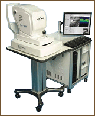
Green laser 532nm Iridex
It has all three delivery devices Viz. Slit lamp, Indirect Ophthalmoscope and endolaser Laser treatment for, retinal holes, tears, lattice degeneration, retinal vascular diseases

Vitrectomy System
It is equipped with 25 G Vitrectomy; Cutting rate of up to 2500 cuts per minute Revolutionary 3-D technology ensuring spectacular results in Vitreo retinal surgeries

Zeiss Visu 160 microscope with BIOM
- Top of the line microscope with XY and motorized zoom
- BIOM and SDI for superior viewing of the retina and the vitreous during surgery – The most advanced wide angle viewing system
- Panoramic view of the retina with excellent resolution and depth perception during surgery
- CCTV with video recording facilities
- All the above state of the art technology ensures excellent results for Vitreoretinal surgeries